Hermetospheres

Experiences with plant life in closed glass containers

, , ,

From transience to permanence

Discover how I learned with one of my hermetospheres what an independent fern gametophyte is.

In a recent post I reported three different bryophytes, that had appeared unintended in the same container. Today, I know I was wrong back then. What I had identified as a liverwort turned out to be an independently propagating fern gametophyte, a thing I didn’t even know exists. Let me explain.

All ‘land plants’, i.e. bryophytes (mosses, liverworts, hornworts), pteridophytes (ferns, lycophytes) and seed plants (gymnosperms, angiosperms), undergo an alternation of generations. This means that their life cycle alternates between a gametophyte and a sporophyte generation. With the exception of bryophytes, the sporophyte generation dominates in all land plants. This is also true of ferns (see the illustration below): what we intuitively identify as a fern plant is the sporophyte. The gametophyte is generally small, short-lived and lacks morphological characters that would allow for species identification (Yoneoka e.a. 2024), and is therefore frequently overlooked. Even in botanical science, the study of diversity of gametophytes has been neglected for many years (Yoneoka e.a. 2024).
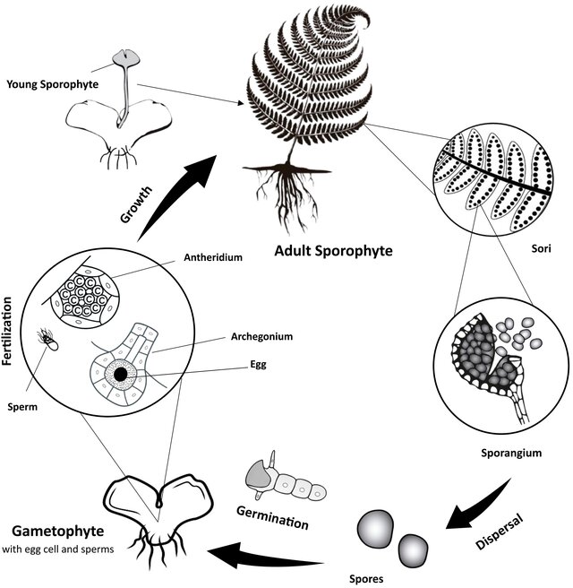
"Figure 2. Simplified view of fern life cycle, illustrating the two generations (sporophyte and gametophyte) and the spores, which represent the main focus of this review. Adult sporophyte leaves produce sori with sporangia full of spores. At maturity, the sporangia shed the spores. Spores germinate and produce the prothalli of the gametophyte. Within the gametophyte, fertilization takes place and a new young sporophyte emerges." (Firoozi e.a. 2025, Figure 2, licenced under CC BY 4.0)
“Figure 2. Simplified view of fern life cycle, illustrating the two generations (sporophyte and gametophyte) and the spores, […]. Adult sporophyte leaves produce sori with sporangia full of spores. At maturity, the sporangia shed the spores. Spores germinate and produce the prothalli of the gametophyte. Within the gametophyte, fertilization takes place and a new young sporophyte emerges.” (Firoozi e.a. 2025, Figure 2, licenced under CC BY 4.0)

The fern gametophyte is also called prothallium or prothallus. Pinson e.a. (2017: fig. 1) give a nice overview on how fern prothalli can look like (see the Figure below). Particularly among tropical, epiphytic fern species, gametophytes with more complex structures and longer lifespans have evolved. These traits are thought to increase the likelihood of two individuals coming into close enough proximity for outcrossing to occur. In three families (Hymenophyllaceae, Polypodiaceae, Pteridaceae), several species have further independently evolved the ability to reproduce asexually via small vegetative propagules called gemmae. These are thought to enhance the likelihood of establishment of fern populations in the canopy, as only one gemma needs be dispersed to give rise to an entirely new population. (Pinson e.a. 2017: 2)
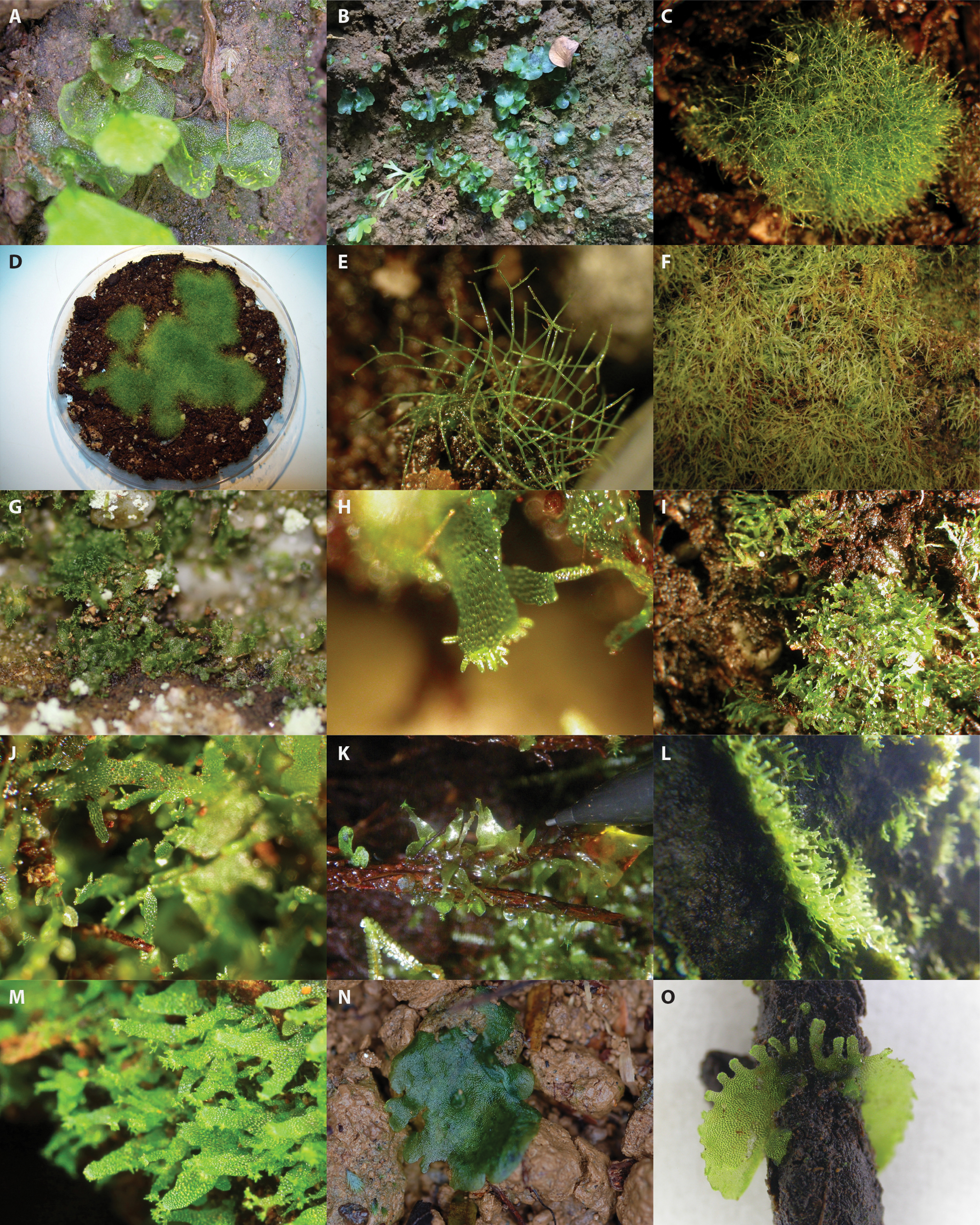
Fig. 1. Photographs illustrating the morphological diversity of fern gametophytes. A, B, Typical cordiform gametophytes (species unknown). The left-hand gametophyte has an emergent young sporophyte slightly out of focus in the foreground. C–E, Crepidomanes intricatum (Hymenophyllaceae). F, Hymenophyllum tayloriae (Hymenophyllaceae). G, Vittaria appalachiana (Pteridaceae). H–J, Hymenophyllum wrightii (Hymenophyllaceae). K, Callistopteris apiifolia (Hymenophyllaceae). L, Vaginularia paradoxa (Pteridaceae). M, Haplopteris heterophylla (Pteridaceae). N, Danaea nodosa (Marattiaceae). O, Anetium citrifolium (Pteridaceae). A, N, and O by J. E. Watkins Jr. B and F by E. Sessa. C–E and H–J by A. Duffy. G by S. Chambers. K and L by J. Nitta. M by C.-W. Chen. Image Source: Pinson e.a. (2017: fig. 1), all rights protected.

One of the most famous examples of an independent fern gametophyte is Vittaria appalachiana. The sporophytes of this species are thought to be extinct due to global cooling during the last glacial period, whereas the associated gametophytes (see the two pictures below) with stronger cold tolerance survived with gemma reproduction around the Appalachian Mountains of North America for more than 10,000 years (Farrar and Mickel 1991, cited in Yoneoka e.a. 2024: 816)
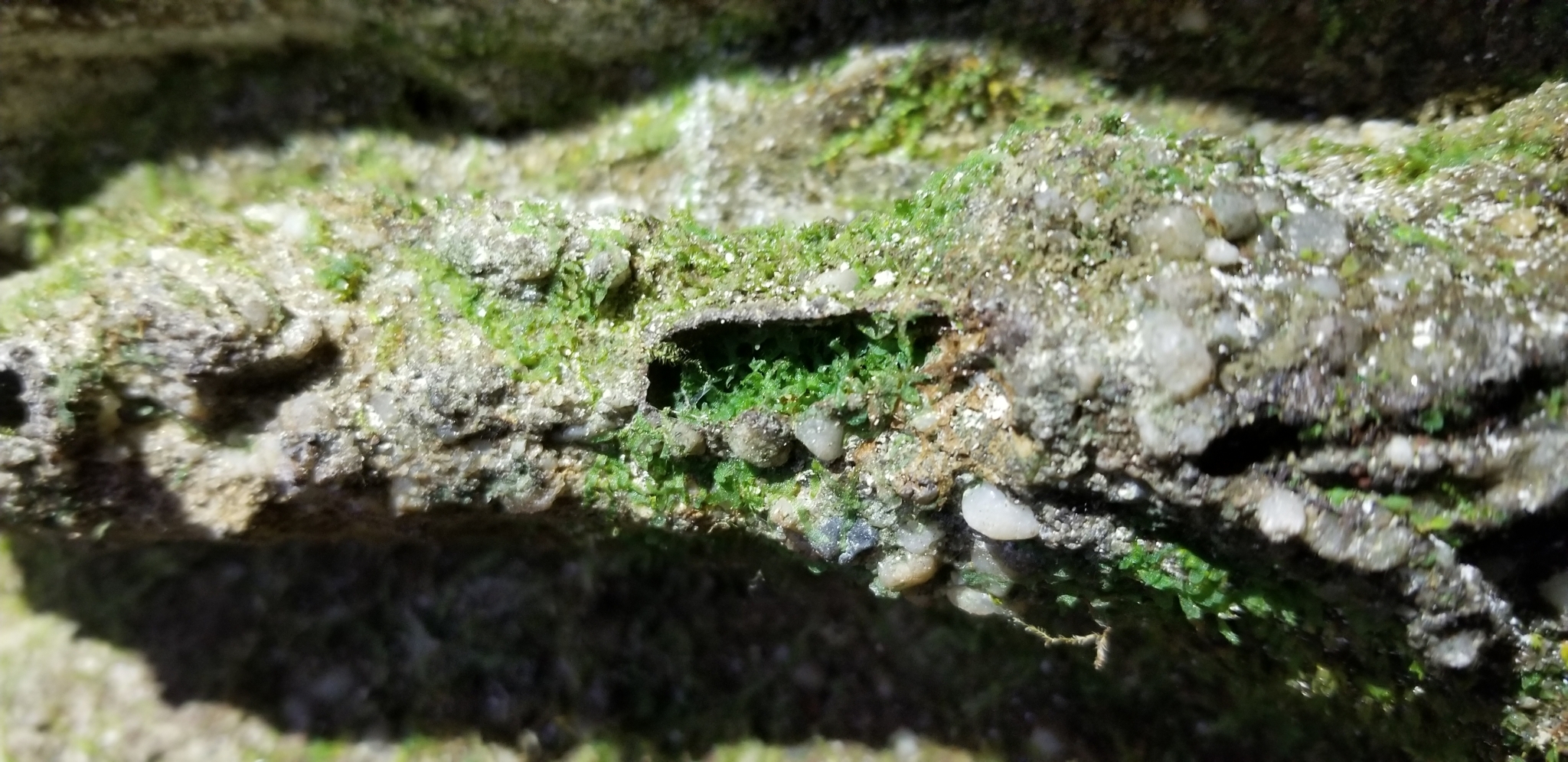
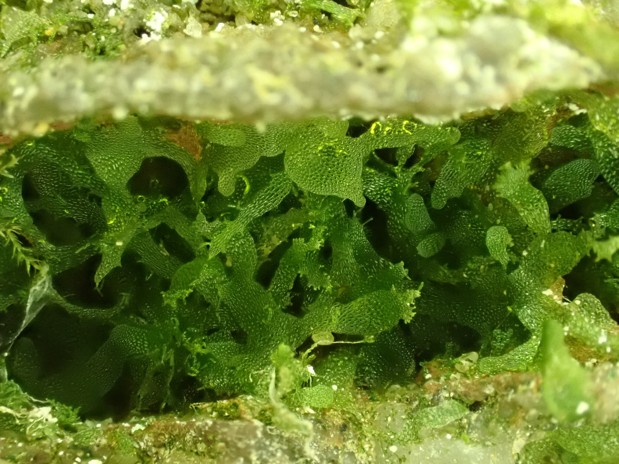
Vittaria appalachiana Farrar & Mickel observed 2023 in Montgomery KY, USA, by Barb Graham (licensed under CC BY-NC 4.0).

Back to my own observation: What I now know is a fern gametophyte appeared ‘out of nowhere’ in one of my closed containers (see picture below).
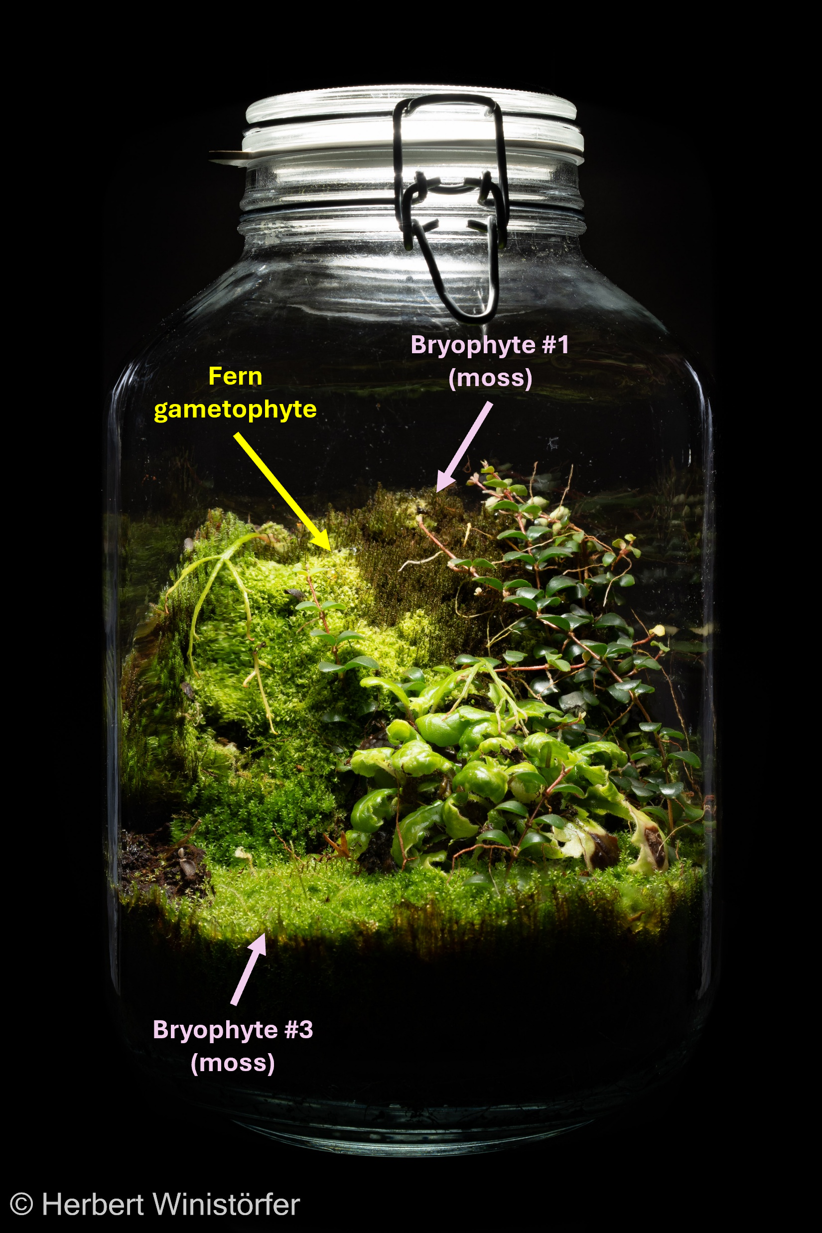
Container of 5 litres with, in addition to three vascular plants, two different mosses (moss #1, moss #3) and an independent fern gametophyte; 29 December 2025, 2 years after onset.

A few small patches were transferred into a sealed glass tray with transparent lid on substrate similar to that I use for my hermetospheres. After 16 weeks, the gametophyte had spread over half of the substrate surface (see picture below).
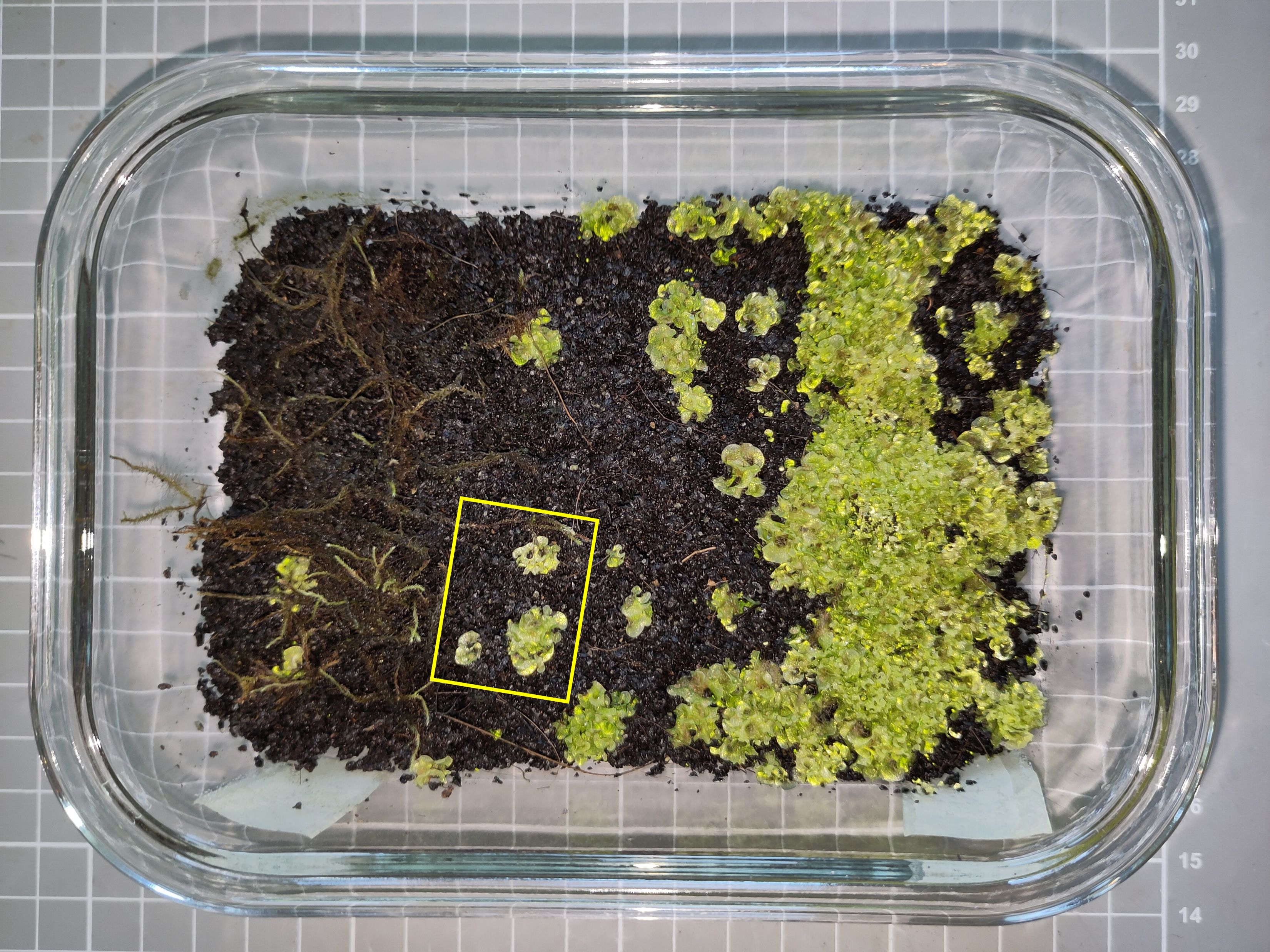
Cultivation of the independent fern gametophyte in a sealed tray on a substrate made of lava sand, cocopeat and earthworm humus; 15 June 2026, yy weeks after inoculation; the rectangle marked in yellow indicates the section enlargd in the following image.
Cultivation of the independent fern gametophyte in a sealed tray on a substrate made of lava sand, cocopeat and earthworm humus; 15 June 2026, 16 weeks after inoculation; the rectangle marked in yellow indicates the section enlargd in the following image.

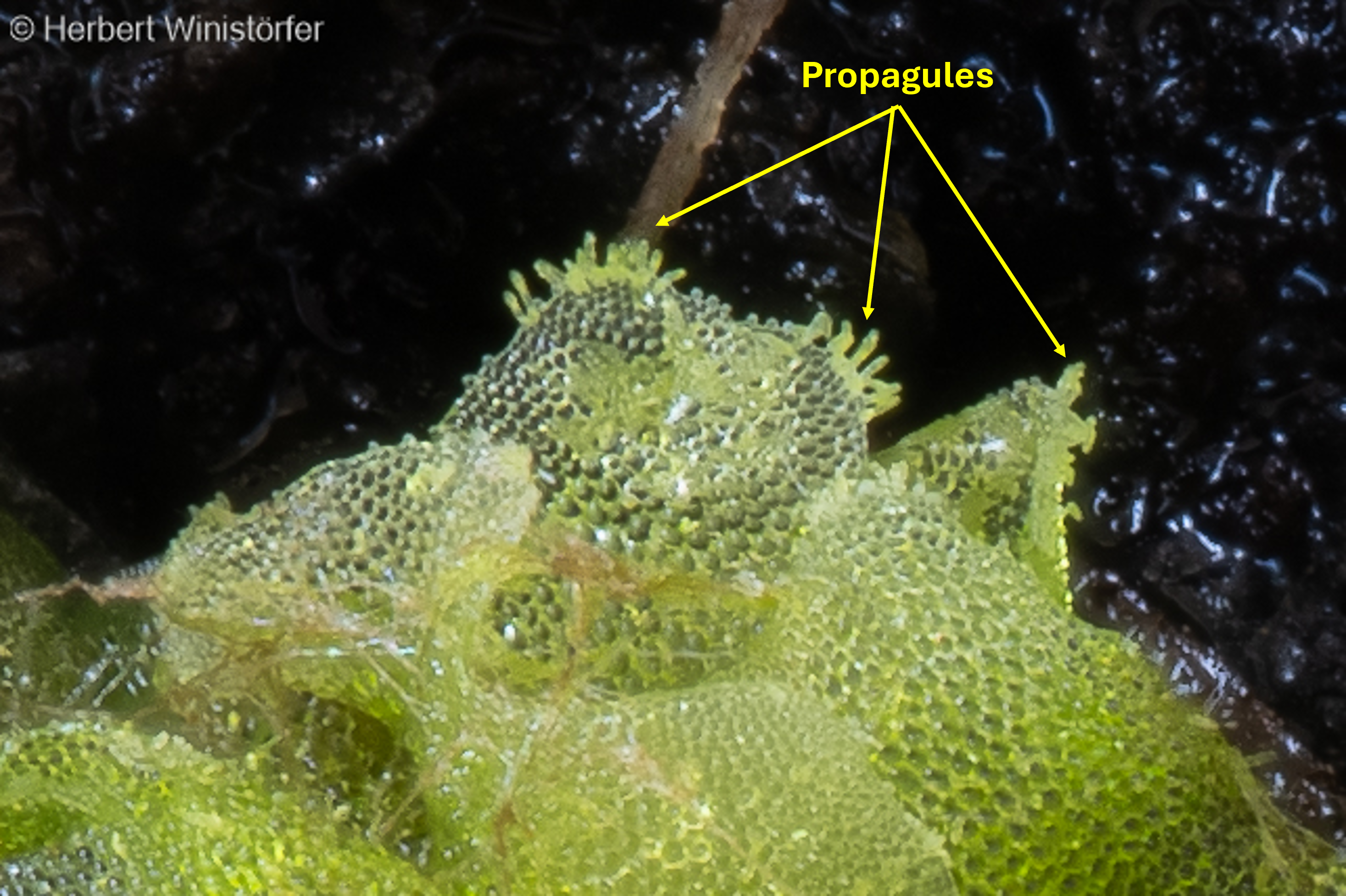
I began to closely examine and document what had been grown. The thallus looked like a single layer of cells, and certain regions were full of growing or mature propagules (see the pictures below). This, I thought, should be enough to identify the plant. A photograph of a product sold by some retailers specialising in plants for terrariums put me on the right track.
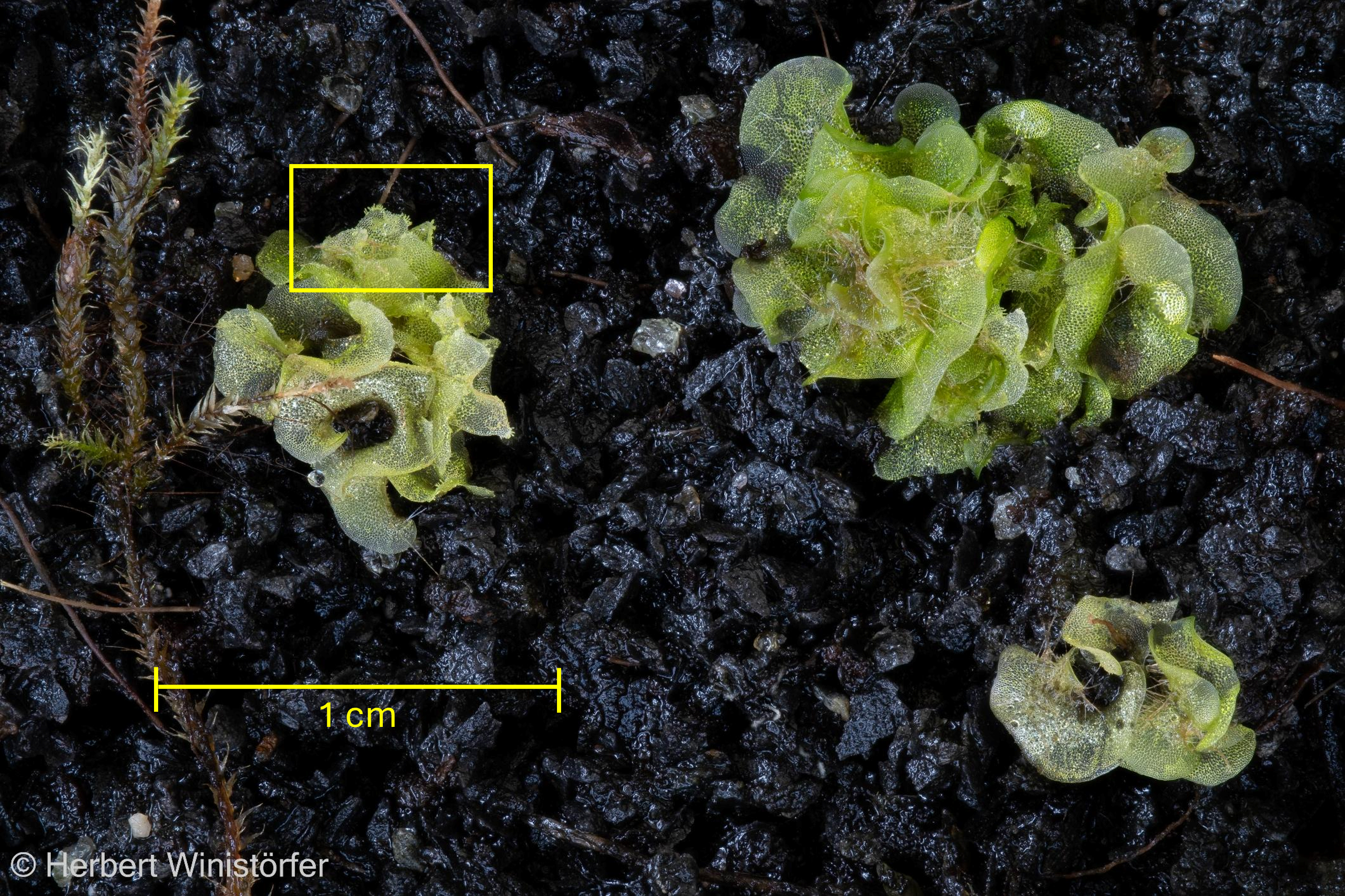
Cultivated fern prothalli grown in a sealed tray on a substrate made of lava sand, cocopeat and earthworm humus; 13 June 2026; the rectangle marked in yellow shows the section that is enlarged in the following image.
Detail from the previous image thar shows the propagules formed at the borders of the prothallium; 13 June 2026.
Detail from the previous image that shows the propagules formed at the borders of the thallus; 13 June 2026.
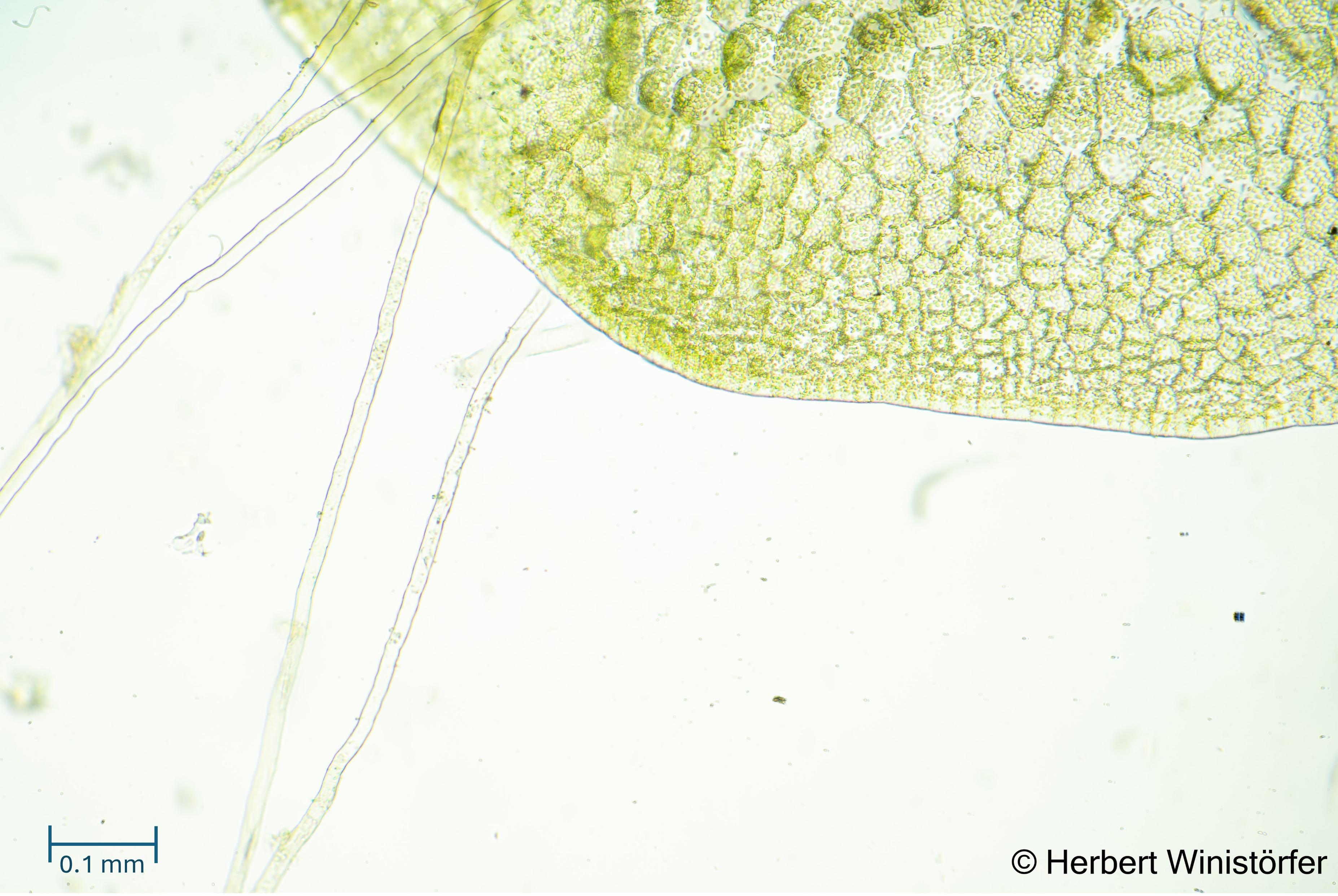
Thallus and rhizoids of the independent gametophyte observed with the transmission light microscope; 20 June 2026; image focus stacked from 76 single frames.
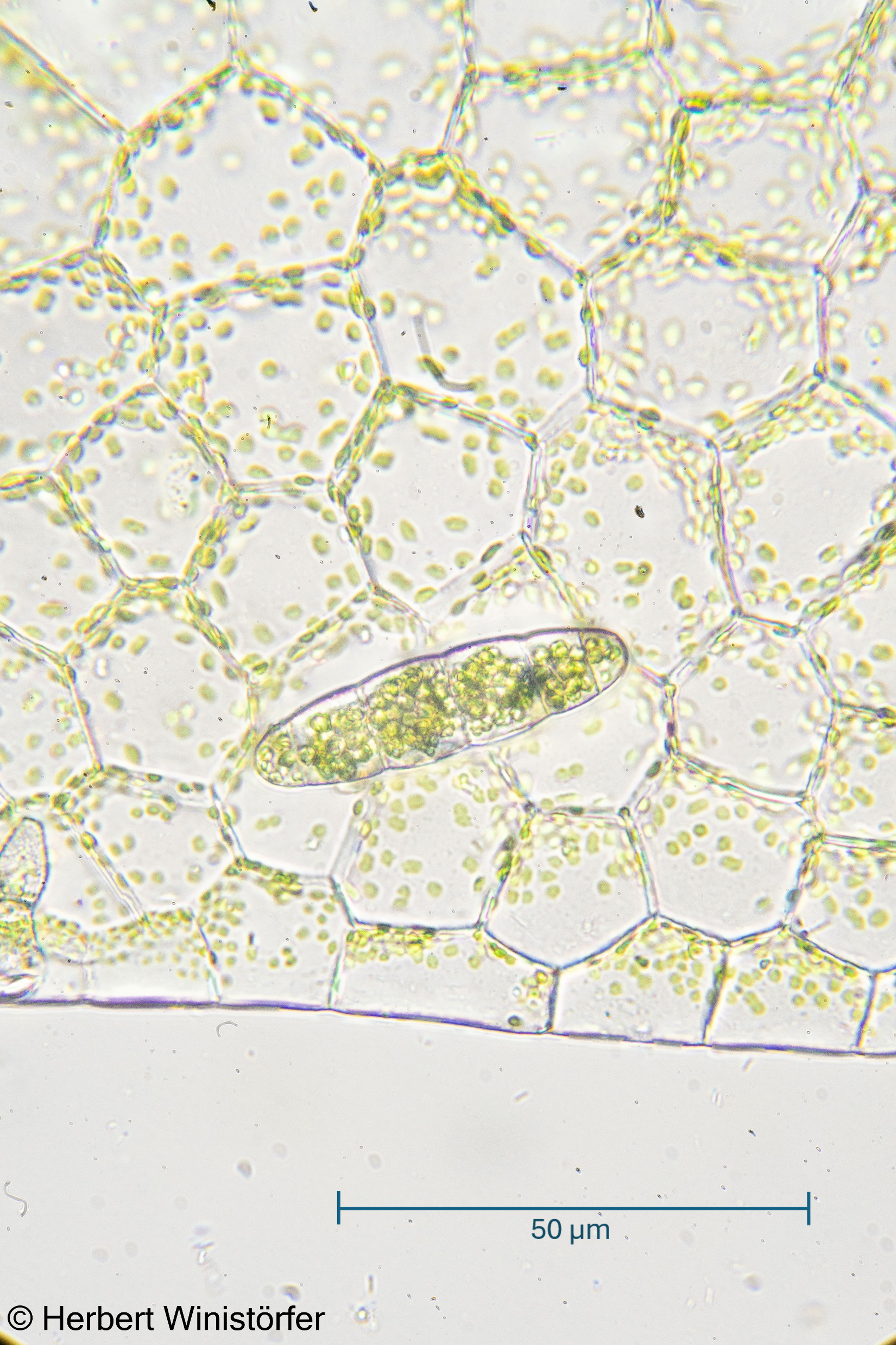
Margin of the thallus and six-celled propagule of the independent gametophyte observed with the transmission light microscope; 21 June 2026; image focus stacked from 93 single frames
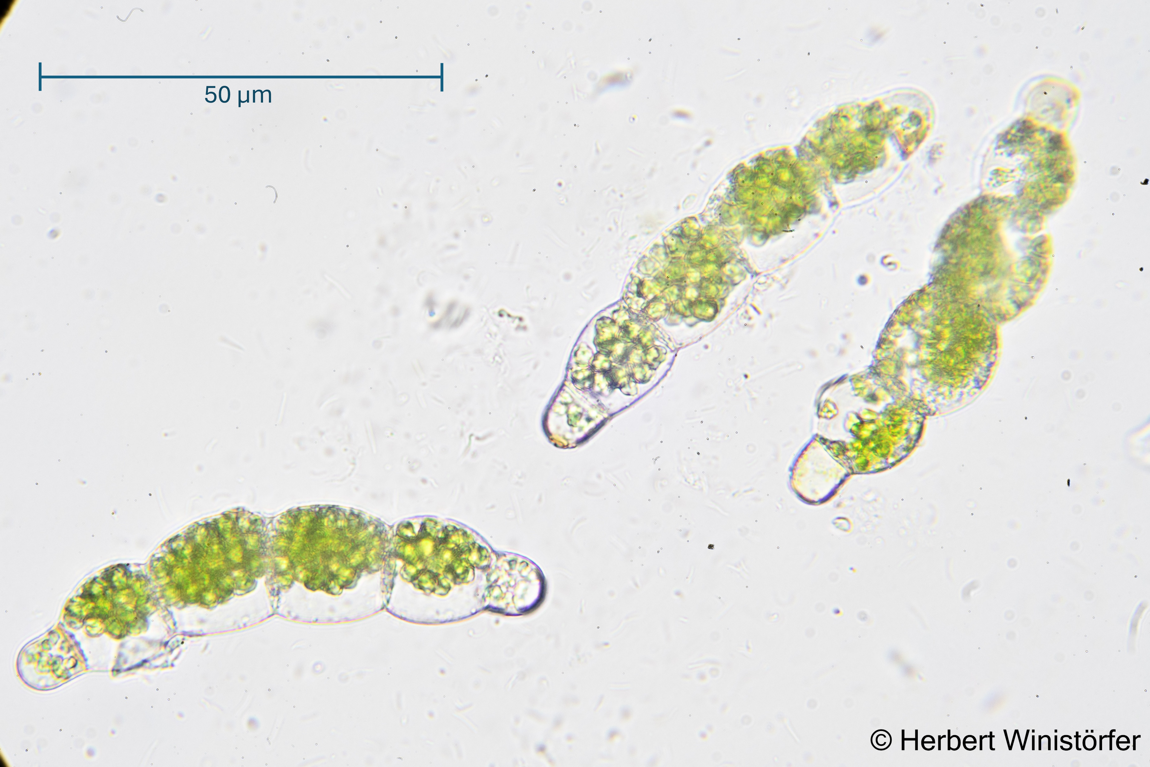
Six-celled propagules of the independet fern gametophyte observed with the transmission light microscope; 15 March 2026; image focus stacked from 2 single frames.

Independent fern gametophytes are sold by some traders under the name of Antrophyum cf. plantagineum as a ‘miniature fern’. A quick check in the GBIF database reveals that the sporophyte of the true A. plantagineum is far from miniature. How the identification of the traded material was established and whether all offerings stem from the same clone, I cannot tell. It is possible that the clone in trade originates from a collection on the Ogasawara Islands, Japan. Murakami e.a. (2021:7) describe occurrences of A. plantagineum as an independent gametophyte from this location. These oceanic islands lie approximately 1,000 km south of central Tokyo and have never been connected to any large landmass. Interestingly, the corresponding sporophyte is not known either from these islands nor from the Japanese mainland. Instead. It is found from India to Sumatera and Jawa to Northwestern Australia. The fact that this product is available in the terrarium trade – and thus grows in the associated greenhouses – could well explain how it managed to find its way into my jar unnoticed. All it takes is a single one of the six-cell propagules pictured above.

Identifying a fern gametophyte without an associated sporophyte can be difficult or even impossible, even for a specialist, particularly when, as in my case, its geographical origin is unknown. In recent years, a new method has led to significant progress in this area: “DNA barcoding, the use of a standard, short region of DNA for species identification (Hebert et al., 2003a), is a powerful method to enable species identification of field‐collected pteridophyte gametophytes. Since this method became available, it has revolutionized the study of pteridophyte ecology by enabling accurate identification of this cryptic sexual life stage over a wide taxonomic range.” (Nitta and Chambers 2022: 2)

Maybe, when in a near future DNA barcoding for fern gametophytes will be affordable for hobbyists, I might want to find out the true identity of my gametophyte. For now, I am happy with what I have learnt so far from my mysterious hitchhiker.

Leave a comment